Battery Microstructure Mapping Is Getting Easier, Less Destructive
A ZEISS expert explains how technologies such as 3D X-ray microscopy, computed tomography, and nanotomography simplify battery microstructure mapping.
February 20, 2023
Herminso Villarraga-Gómez, ZEISS
As humanity’s future energy sustainability only grows in importance, the electric vehicle industry is becoming the largest market for high-performance rechargeable batteries to the point in which nearly every major automaker has announced a transition away from gas-powered cars to electric vehicles in the next ten to thirty years.
Still, further advances in cell design and manufacturing processes are necessary to improve energy density, capacity, energy storage retention, cycle performance, lifespan, and safety of rechargeable batteries.
Significant advances are continually occurring in rechargeable battery technologies, but battery cell manufacturers still face quality and process control challenges when attempting to non-destructively map the microstructure of battery electrodes, their inhomogeneities, and their effect on battery ageing and performance degradation.
As such, advanced technologies such as 3D X-ray microscopy will figure largely in advancing new battery development. While lithium-ion batteries (LIBs) have become an important driver in the transition to a carbon-free future, methods similar to those described here can certainly be applied to other battery systems, e.g. solid-state batteries built with other emerging energy materials.
Testing without destroying
Of the various techniques that can be used to image battery components (e.g. visible light, ultrasound, X-rays, electron beam, neutron beam, etc.), X-ray imaging is the only technique that can non-destructively inspect the interior of battery cells across multiple length scales—from centimeters (battery packs and cell-level analysis) to submillimeter volumes (material microstructure-level analysis)—and capture relevant structural details with spatial resolutions ranging from several tens of micrometers down to tens of nanometers.
Although using 3D X-ray imaging technologies to inspect LIBs is relatively recent, it has rapidly gained ground. Spatial resolution requirements and sample size generally set the limits of X-ray imaging setups. While traditional flat-panel-based high-energy CTs are suitable for inspecting large battery packs and battery assemblies, they generally lack the resolution needed to inspect fine-scale features below 10 μm.
With the addition of optical lenses after X-ray detection, 3D X-ray microscopes (XRMs) can overcome these challenges. However, due to power and energy limitations, higher spatial resolutions will limit the sample’s field of view. XRMs are ideal for inspecting small battery cells and components in the 0.1 to 200-mm range, including individual electrodes and separators.
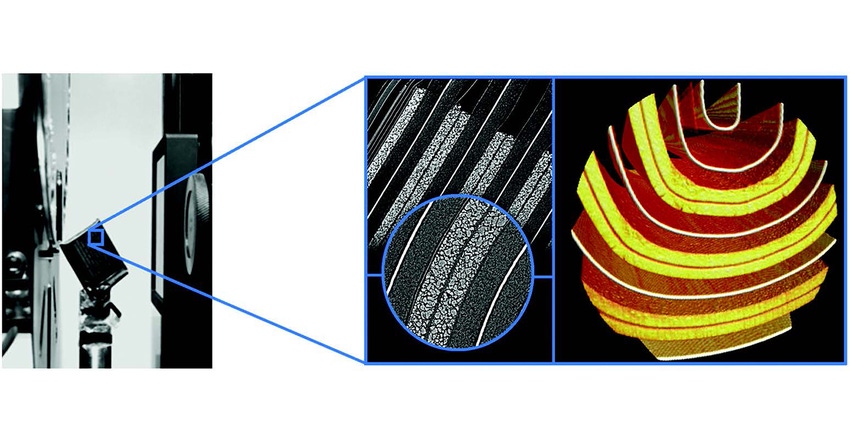
Due to the non-destructive nature of X-ray microscopy, the interior arrangements of complete battery cells can be imaged without cutting into the cells. This allows engineers and researchers to observe the arrangement of internal features of intact batteries, such as layer stacking alignment, electrode thickness distribution, microscopic inhomogeneities in electrode materials, and the existence of electrode defects from the production processes.
An example
In smaller-scale cells, such as pouch cell batteries used in consumer electronics (e.g. smart watches and cell phones), detailed views of the electrode microstructure can be observed.
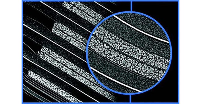
The illustration at the top of this article shows 3D XRM images of pouch cell batteries. The center image (enlarged above) shows the detailed microstructure of the cathode layer—those thick, bright bands—and the associated particles that make up the layer (as seen in the inset).
Cracking defects can also be seen in the cathode layers due to electrode bending in pouch cell packaging, as shown in the image of a commercial cell phone battery below. These types of microstructure defects are generally associated with volume expansion and contraction of active material particles during lithiation/delithiation cycles, mechanical stresses, thermal fatigue, or other saturation conditions leading to charge capacity deterioration and power fading.
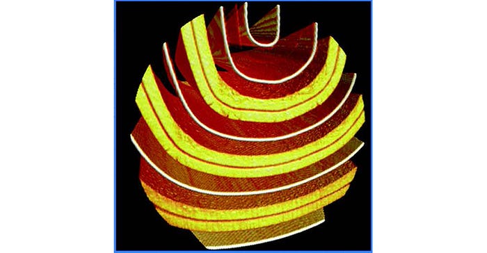
The non-destructive imaging capability of X-rays permit investigating and understanding energy efficiency, the effects of ageing, and battery cell degradation and failure after multiple charge/discharge cycles, to aid in the design and quality assessment of battery cells.
Due to the multiscale nature of rechargeable batteries, with relevant feature dimensions ranging from nanometers to centimeters, the use of correlative imaging workflows that include imaging methods other than X-rays (e.g. electron microscopy) may be beneficial, and in some cases the mere use of correlative X-ray imaging may be sufficient.
In conclusion
Workflows that combine computed tomography and 3D X-ray microscopy can generate a detailed three-dimensional visualization of battery cell interiors and assemblies—without destroying them—enabling the study of their internal structure before and after charging/discharging cycles. These imaging workflows can be run independently or complementary to other multiscale correlative microscopy evaluations and provide valuable insights into the inner workings of battery systems at multiple length scales.
Understanding battery systems through X-ray imaging can speed development time, increase cost efficiency, and simplify failure analysis and quality inspection of lithium-ion batteries (LIBs) and other cells built with emerging energy materials.
Herminso Villarraga Gomez is Product Manager for X-ray Quality Solutions and Global Sector Marketing for 3D X-ray Microscopy at ZEISS. For further reference, see Villarraga Gómez et al., Assessing rechargeable batteries with 3D X-ray microscopy, computed tomography, and nanotomography, Nondestructive Testing and Evaluation, 37:5, 519-535, 2022.
You May Also Like
